The complex and highly variable anatomy of the equine endodontic system, particularly in cheek teeth, represents a major diagnostic challenge in equine dentistry. Multiple pulp horns, age-related morphological changes, and secondary dentin deposition complicate both the assessment of endodontic status and the planning of appropriate therapeutic interventions. Advanced imaging techniques have significantly improved the understanding of equine dental anatomy. Micro-computed tomography (micro-CT) provides highly detailed three-dimensional insights into pulp morphology and interconnections and is therefore an invaluable tool in research. In clinical settings, however, evaluation of pulp vitality is of particular importance for treatment decision-making. Magnetic resonance imaging (MRI) has been demonstrated to provide the most reliable information on pulp vitality (Gerlach et al.., 2013). Nevertheless, due to limited availability, high costs, and logistical constraints, both micro-CT and MRI are rarely applicable in routine equine practice. Consequently, clinicians must rely on more accessible imaging modalities. Following thorough clinical and oroscopic examination, radiography and computed tomography (CT) represent the cornerstone of endodontic diagnostics in horses. CT allows for detailed three-dimensional assessment of dental structures and is particularly useful for identifying apical infections, pulpar changes, and associated sinus involvement. Despite these advantages, CT is typically not available intraoperatively. Conventional radiography therefore remains indispensable during endodontic procedures. It provides essential real-time guidance for instrumentation and allows the detection of communications between the endodontic system and surrounding structures, such as fistulae or paranasal sinuses.Radiographic contrast studies further enhance diagnostic capabilities by visualizing these communications. Barium sulfate-containing contrast media are commonly used to delineate pathways between the pulp system and adjacent cavities. Accurate endodontic instrumentation relies heavily on radiographic control. Due to the inherent limitations of two-dimensional imaging, multiple projections at different angles are often required to precisely determine the position and depth of instruments, such as endodontic files, and to avoid procedural errors.
Take-home messages
• The anatomical complexity of the equine endodontic system necessitates a multimodal imaging approach.
• Micro-CT and MRI provide superior anatomical and functional information but are currently limited to research or selected clinical cases.
• CT is the most informative clinical imaging modality for diagnosing endodontic pathology, particularly in relation to apical disease and sinus involvement.
• Conventional radiography remains the key tool for intraoperative guidance and endodontic instrumentation. (Image1)
• Contrast radiography is a valuable adjunct for identifying communications with fistulae and adjacent cavities. (Image 2)
• Multiple radiographic projections are essential to compensate for the limitations of two-dimensional imaging and to ensure accurate instrument placement.
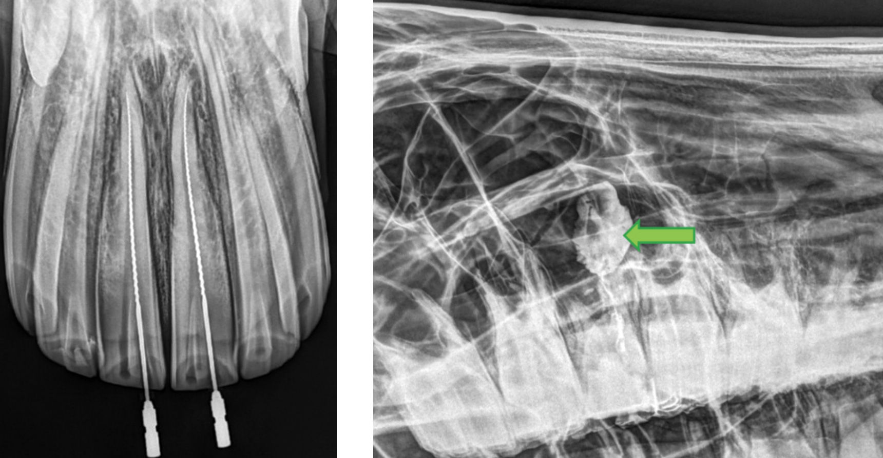
Image 1: Radiographic
Image 2: Contrast study confirm communication control of file positioning of 209 (Pulphorn 1) with rostral maxillary sinus
References
1. Gerlach K, Ludewig E, Brehm W, Gerhards H, Delling U. Magnetic resonance imaging of pulp in normal and diseased equine cheek teeth. Vet Radiol Ultrasound. 2013 Jan-Feb;54(1):48-53. doi: 10.1111/j.1740-8261.2012.01971.x. Epub 2012 Sep 25. PMID: 23006129.
2. Townsend NB, Hawkes CS, Rex R, et al.. Investigation of the sensitivity and specificity of radiological signs for diagnosis of periapical infection of equine cheek teeth. Equine Vet J 2011;43(2):170–8.
3. du Toit N. Aetiology and diagnosis of periapical dental disease in equids. Equine Vet Educ 2011;23(11):559–61.
4. Selberg K, Easley JT. Advanced imaging in equine dental disease. Vet Clin North Am Equine Pract. 2013 Aug;29(2):397-409, vi. doi: 10.1016/j.cveq.2013.04.009. Epub 2013 May 28. PMID: 23915666.
5. Stoll M. Verfeinerung der Röntgendiagnostik am Pferdekopf mit Hilfe von Kontrastmitteleinsatz IGFP Kongress Proceedings 2015
